East Delhi's Most Trusted and accurate ultrasound

Ultrasounds with Accuracy and Affordability - Call Now!
NABH accredited, 35 year Old Hospital and Diagnostics Centre for you


Latest and Best Technology
Best in class Equipment with
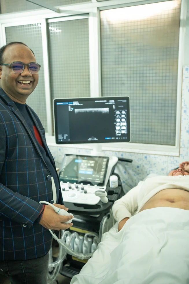
SAMSUNG V7 Ultrasound
Global Standard Reporting
Advanced reporting system with universally recognized standard, providing comprehensive and reliable results tailored to your condition.
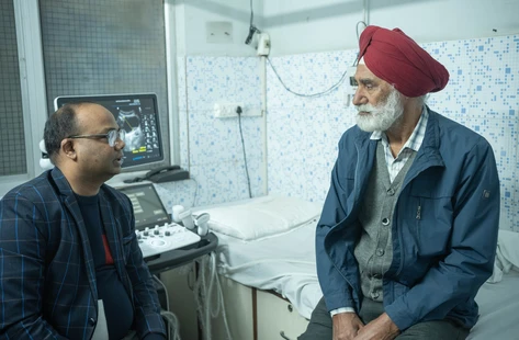
Experienced Radiologist
who brings years of expertise to interpret your scans, with meticulous analysis and accurate diagnostics
Level II ultrasound
Ensure the well being of your young one during pregnancy with an accurate and precise Level II Ultrasound
Rapid Results, Reliable Diagnosis
Convenience of swift ultrasound tests without compromising on accuracy and reliability!

35 Years of Trust
5 Lakh + Happy Patients over three decades speak for us!
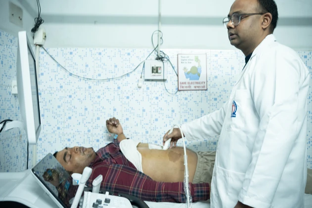
Abdominal Ultrasound
Whole Abdomen ultrasound scan is conducted to diagnose conditions such as gallstones, liver disease, appendicitis, or abdominal tumors, helping to identify the underlying cause of symptoms.
Provides a comprehensive view of abdominal organs.
Post Void Residual Urine Test Evaluates bladder function after urination to assess urinary retention.
Combines abdominal and pelvic imaging to investigate potential issues in both areas.
Incorporates obstetric ultrasound to monitor fetal development alongside abdominal examination.
Liver Ultrasound
A liver fibroscan is like a health checkup for your liver, but without the need for surgery. It’s a simple test that uses a specialized ultrasound machine to assess how flexible or stiff your liver is. This information helps doctors figure out if there’s any scarring or damage in your liver, which can be caused by conditions like hepatitis or fatty liver disease. During the test, a small probe is placed on your skin over your liver, and it sends sound waves through your liver to measure its stiffness. This process is quick, painless, and doesn’t require any needles or incisions.
Non-invasive diagnostic test utilized to evaluate liver health and detect the presence and extent of liver fibrosis, using a specialized fibroscan machine. art, lungs, and mediastinum.
Obstetric/Pregnancy Ultrasound
Obstetric ultrasound is essential for monitoring fetal growth, ensuring proper development, and detecting any abnormalities early, or an Anomaly Scan in pregnancy and providing crucial information for prenatal care. Scans like Level ii scan are instrumental during pregnancy and we ensure you get the most comprehensive results.
Screens for chromosomal abnormalities, such as Down syndrome, in early pregnancy.
Utilized during pregnancy to monitor fetal growth, position, and well-being.
Assesses fetal health by evaluating fetal movements, muscle tone, breathing, and amniotic fluid levels.
Provides detailed anatomical assessment of the fetus, often performed around 18-22 weeks of pregnancy.
Echo tests
At EDMC our expert Medical team conducts two Echo Tests. We provide detailed, two-dimensional images of your heart’s structure and function. From assessing chambers and valves to detecting potential abnormalities, our 2D Echo tests offers a comprehensive evaluation to guide your healthcare decisions.
A 2D echocardiogram, also known as a two-dimensional echocardiogram, is a non-invasive imaging test that provides detailed images of the heart’s structure and function. Using high-frequency sound waves (ultrasound), the test allows healthcare professionals to visualize the heart’s chambers, valves, walls, and blood vessels in real-time. 2D echocardiograms are commonly used to diagnose various heart conditions such as valve disorders, congenital heart defects, cardiomyopathy, and abnormalities in the heart’s size or shape.
A stress echocardiogram, or stress echo, is a specialized imaging test that evaluates the heart’s function under stress conditions. It combines echocardiography with either exercise (treadmill or stationary bike) or medication to induce stress on the heart. During the test, ultrasound images of the heart are obtained at rest and then again immediately after stress is induced. This dynamic assessment allows healthcare professionals to observe how well the heart responds to increased demands for oxygen-rich blood during physical exertion or pharmacological stress.
Pelvic Ultrasound
Pelvic ultrasound, including transvaginal sonography, offers non-invasive imaging of reproductive organs like the uterus and ovaries. This technique aids in diagnosing conditions such as ovarian cysts and fibroids
Involves inserting a probe into the vagina to visualize pelvic organs like the uterus, ovaries, and cervix.
Examines small structures like breasts, or inguinal area for abnormalities.
Chest Ultrasound
Chest Ultrasound help in diagnosing thoracic region with detailed imaging of Lungs, hearts and Surrounding issue and can detect cardiac anomalies, lung nodules and pleural effusions.
Examines structures within the chest cavity, including the heart, lungs, and mediastinum.
Offers combined chest and abdominal imaging for comprehensive evaluation, useful in diagnosing conditions affecting both areas.
Color Doppler Ultrasound
Color Doppler ultrasound assesses blood flow in arteries and veins & During prenatal care, Doppler ultrasound or Doppler sonography, assess blood flow in the placenta, umbilical cord, and fetal vessels & can detect any potential abnormalities early on
assesses blood flow in the kidneys and renal arteries, aiding in the diagnosis of conditions such as renal artery stenosis or kidney dysfunctionngs, and mediastinum.
Evaluates blood flow in the carotid arteries supplying the brain, assisting in assessing the risk of stroke, detecting carotid artery disease, and guiding treatment to prevent stroke and other cerebrovascular events
Assesses blood flow in the arteries of Single and Both Limbs, aiding in the diagnosis of peripheral artery disease (PAD), deep vein thrombosis (DVT), or other vascular conditions affecting circulation in the lower extremitiesd mediastinum.
Assesses blood flow in the umbilical artery of the fetus, aiding in the assessment of fetal well-being and monitoring placental function, particularly in high-risk pregnancies
Evaluates blood flow in the testicular arteries and veins, aiding in the diagnosis of conditions such as testicular torsion, varicocele, or epididymitis, assisting in the evaluation of testicular pain, swelling, or infertility issues
Specialized Ultrasound Procedures
For procedures such as guided fine-needle aspiration cytology (FNAC) and the insertion of a pigtail catheter for drainage, can be facilitated by ultrasound guidance, ensuring precision and minimizing risks. Moreover, in cases of liver abscesses, ultrasound-guided procedures aid in accurate diagnosis and targeted treatment, enhancing patient outcomes.
Focuses on specific anatomical regions, facilitating the diagnosis of conditions affecting these areas
Guides needle insertion for draining liver abscesses, aiding in the treatment of liver infections.
Assesses structures within the skull, including the brain and cerebral blood vessels.
Involves inserting a drainage catheter under ultrasound guidance, often used in the management of fluid collections
Utilizing ultrasound technology, therapeutic tapping delivers targeted pain relief and promotes healing by stimulating circulation and reducing inflammation in affected areas.
Our diagnostic tapping procedure employs ultrasound imaging to accurately diagnose internal conditions and injuries, guiding treatment decisions with precision and efficiency.
Guides fine needle aspiration for tissue sampling, aiding in the diagnosis of suspicious masses or lesions.
Let's Understand Ultrasound Tests!
What is an ultrasound?
Ultrasound Test is a medical test that uses high-frequency sound waves to create images of the inside of the body. It helps doctors see organs, tissues, and structures in real time and helps in diagnosing various issues in body.
How does an ultrasound work?
During an ultrasound, a small device called a transducer sends sound waves into the body. These waves bounce off organs and tissues, creating echoes. The transducer picks up these echoes and turns them into images on a screen.
Which diagnostic Tests can I get done at EDMC?
We are one of the best and most trusted diagnostic centers of East Delhi! For Urology we offer imaging studies (such as ultrasound and CT scans), urine tests, cystoscopy, urodynamic testing, and prostate-specific antigen (PSA) testing. These help us accurately diagnose and develop personalized treatment plans.
Do I need to prepare for an ultrasound?
Preparation for an ultrasound depends on the type of exam. For some, you might need to fast for a few hours, while for others, you might need to drink water and have a full bladder. Your healthcare provider will give you specific instructions.
Who performs ultrasounds?
Ultrasounds are typically performed by trained technologists or sonographers who specialize in using ultrasound equipment to create images. Doctors then interpret these images to make diagnoses.
Why are ultrasounds used?
Ultrasounds are used for various reasons, like checking the growth of a baby during pregnancy, examining organs like the heart, liver, or kidneys, detecting abnormalities, guiding medical procedures, and more.
Are ultrasounds safe?
Yes, ultrasounds are considered safe because they use sound waves instead of radiation. There are no known risks to the patient during a standard ultrasound procedure.
Do ultrasounds hurt?
No, ultrasounds are painless. You may feel some pressure from the transducer as it’s moved over your skin, but it’s not uncomfortable.
Are there different types of ultrasounds?
Yes, there are different types of ultrasounds based on the area of the body being examined. For instance, there are abdominal ultrasounds, pelvic ultrasounds, fetal ultrasounds during pregnancy, and more.
How long does an ultrasound take?
The duration of an ultrasound can vary depending on the type of exam and what’s being examined. Generally, an ultrasound takes between 20 to 60 minutes.
Hear from our patients